Equipment
Microscopes
Confocal (point scanning)
M24 - LSM780 Axio Imager (upright)
M27 - LSM880 Axio Observer (inverted) with Airyscan/Airyscan fast
M33 - LSM880 Axio Observer (inverted) with Airyscan/Airyscan fast and incubation and 40x/1.2W, Glycerol AutoCorr objective
M35 – LSM800 Examiner Z1 (upright) with incubator
M36 – LSM800 Axio Observer Z1 (inverted) with incubator
M37 – LSM800 Axio Imager (upright)
M38 – SP8 DIVE confocal (inverted) with multiphoton lasers and incubation
M41 – LSM980 Axio Observer (inverted) with Airyscan 2 and incubation
M42 – LSM980 Axio Observer (inverted)
M48 – LSM800 / Vertical
M53 – LSM980 Axio Observer (inverted) with Airyscan 2 and incubation
Confocal (spinning disk)
M01 - Spinning Disk Confocal Axio Observer (inverted)
M40 - Spinning Disk Confocal Olympus (inverted) with incubation
M44 - Spinning Disk Confocal Olympus (inverted) with incubation/Superresolution (SoRa)
M50 - Spinning Disk Confocal Olympus (inverted) with incubation and FRAP unit
M54 - Spinning disk Confocal Olympus (inverted) with incubation and FRAP unit
Light Sheet
M32 – Zeiss Z1 Lightsheet Microscope
M45 - Viventis LS1 light sheet
M55 - AxL Cleared Tissue Light Sheet
TIRF/SMLM
M46 - Elyra7 with lattice SIM2
Widefield
M02 - Axio Imager.Z2 (upright) with sCMOS camera
M05 - Axio Imager.Z2 (upright) with sCMOS camera, Axiocam colour camera and Apotome2
M06 - Axioobserver Z1 (inverted) mit sCMOS camera
M16 - Axioobserver Z1 (inverted) with sCMOS camera
M17 - Fluorescence Stereomicrocope
M34 - Discoverer 7 fully automated live cell imaging system
M39 - Discoverer 7 fully automated live cell imaging system
FLIM
M47 - FLIM - Picoquant Fluorescent Lifetime Imaging Microscope
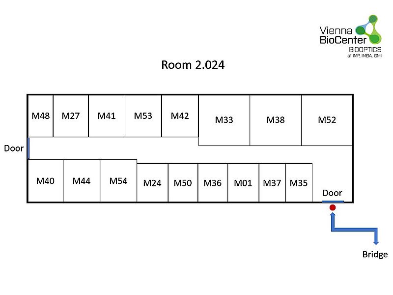
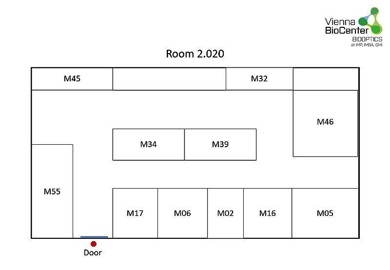
Floorplans Microscopy Rooms