
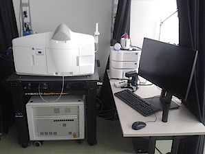
M32 – Zeiss Z1 Lightsheet Microscope
System Description
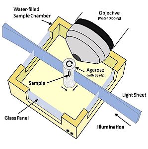
Lightsheet microscopy is the method of choice for delicate living samples and for large cleared samples. A sheet-shaped light beam is used for exciting a single plane of the sample. The fluorescent light from the excited plane is imaged by perpendicular, camera based detection. This results in optical sectioning with low-dosage excitation and next to no phototoxic stress for living samples. Only in-focus volumes that are actually imaged, are illuminated. Using long-distance objectives and fast z-scanning, large cleared samples (up to 1x1x1 cm) can be acquired in short time.
The M32 Zeiss Z1https://www.zeiss.com/microscopy/int/light/lightsheet-z-1.html lightsheet microscope allows for bidirectional excitation, sample rotation, tile scanning, z-scanning and time-lapse imaging. Dual cameras provide fast acquisition for two channels, although with intermittent filter changes, more colors can be acquired easily. An optical zoom ranges from 0.36x to 2.5 x, although reasonable beam shapes (edges not thicker than two times the beam waist thickness) require a zoom of at least 0.71. Samples are mounted on a vertical, hanging support, dipped into the medium and illuminated and imaged horizontally. Scanning perpendicular to the light sheet plane is done by moving the sample through the light sheet. 3D scans are limited to a maximum speed of one stack per second.
The best achievable resolving power along the z-axis of the detection objective is about 2µm (20x/1.0 objective at optical zoom 2.5), providing cellular and sometimes sub-cellular resolution. For cleared samples, proper refractive index matching of embedding medium and imaging medium to the objective is of paramount importance. See the list of available objectives below.
The system is able to run multidimensional experiments including any combination of the following:
· multiple positions
· multiple wavelengths
· z-Stacks, time-lapse
· multiple rotational views
· dual side illumination
· pivot scanning
· incubation (temperature, CO2 bubbling) with 5x dry objective or 10x / 20x water objectives only
Illumination
White Light LED
Laser for Lightsheet
405nm 20mW
445nm 25mW
488nm 50mW
514nm 20mW
561nm 50mW
638nm 60mW
Beamsplitter & Filters
The 5 Beamsplitters divide the emission light into two channels, each of which is detected by a PCO edge sCMOS camera. The beamsplitters are associated with fixed emission filters, for camera one and camera two, respectively.
Set1 - Standard
Beamsplitter 1 (LP 490): cam1 BP [420-470] nm / cam2 BP 505-545 nm
Beamsplitter 2 (LP 510): cam1 BP [420-470] nm / cam2 BP 575-615 nm
Beamsplitter 3 (LP 560): cam1 BP [505-545] nm / cam2 BP 575-615 nm
Beamsplitter 4 (LP 560): cam1 BP [505-545] nm / cam2 LP 585 nm
Beamsplitter 5 (LP 560): cam1 BP [505-545] nm / cam2 LP 660 nm
Set2 - on request
The 5 Beamsplitters divide the emission light into two channels, each of which is detected by a PCO edge sCMOS camera. The beamsplitters are associated with fixed emission filters, for camera one and camera two, respectively.
Beamsplitter 1 (mirror): cam1 none / cam2 none
Beamsplitter 2 (LP 510): cam1 BP [420-470] nm / cam2 BP 525-545 nm
Beamsplitter 3 (LP 510): cam1 BP [460-500] nm / cam2 BP 525-565 nm
Beamsplitter 4 (LP 510): cam1 BP [460-500] nm / cam2 BP 575-615 nm
Beamsplitter 5 (LP 510): cam1 BP [460-500] nm / cam2 LP 585 nm
Beamsplitter 6 (LP 560): cam1 BP [505-545] nm / cam2 LP 660 nm
Excitation Objectives
5x/0.1 (for use with 5x or 10x detection objective)
10x/0.2 (for use with 20x detection objectives)
5x/0.1foc (for use with 5x or 10x detection objective)
10x/0.2foc (for use with 20x detection objectives)
Detection Objectives
5x/0.16: EC-Plan Neofluar 5x/0.16: 5x/0.16 nd=1 WD= 5.1 mm
5x/0.16: foc EC-Plan Neofluar 5x/0.16: 5x/0.16 nd=1 WD= 10.5 mm
10x/0.5: W Plan-Apochromat 10x/0,5 nd=1,33 WD=3.7 mm
20x/1.0: W Plan-Apochromat 20x/1.0 Corr DIC; nd=1.33 WD=2.4 mm
20x/1.0: Clr Plan-Apochromat 20x/1.0 Corr nd=1.38 WD=5.6 mm
20x/1.0: Clr Plan-Neofluar 20x/1.0 Corr nd=1.45 WD=5.6 mm
20x/1.53: Clr Plan-Neofluar 20x/1.0 Corr DIC; nd=1.53 WD= 6.4mm
Imaging Chambers
The following chambers are available:
Water chamber (n = 1.33) to be used with 5×, 10×, 20× and 40× water immersion lenses
20× Clearing chamber (n = 1.35 – 1.58) to be used with Clr 20× immersion lenses
Large sample chamber (n = 1.33 – 1.58) to be used with 5× foc objective
Cameras
PCO Edge 5.5m sCMOS cameras (2x): 6,5 µm/pixel, 1920 x 1920 pixels
Additional Devices
Incubation unit with heating (by a peltier element) and CO2 (bubbling in the imaging medium)